Spinal Sclerosis Homeopathic Treatment
Degenerative scoliosis, also known as adult onset scoliosis, describes a side-to-side curvature of the spine caused by degeneration of the facet joints and intervertebral discs which are the moving parts of the spine. This degeneration and resulting spinal asymmetry can occur slowly over time as a person ages. This is a completely different cause of scoliosis from the standard adolescent onset scoliosis.
If degenerative scoliosis becomes symptomatic, pain can range from a dull back ache to excruciating sensations that shoot down the leg, commonly referred to as sciatica, and make walking difficult or impossible.
How Degenerative Scoliosis Develops
When healthy, facet joints are like hinges that help the spine bend smoothly, and intervertebral discs are like cushions that absorb shock between vertebral bones. Everyone experiences natural degeneration of these joints and discs due to aging—the same processes that cause osteoarthritis and degenerative disc disease—but for some people these degenerative processes are accelerated and/or cause more symptoms.
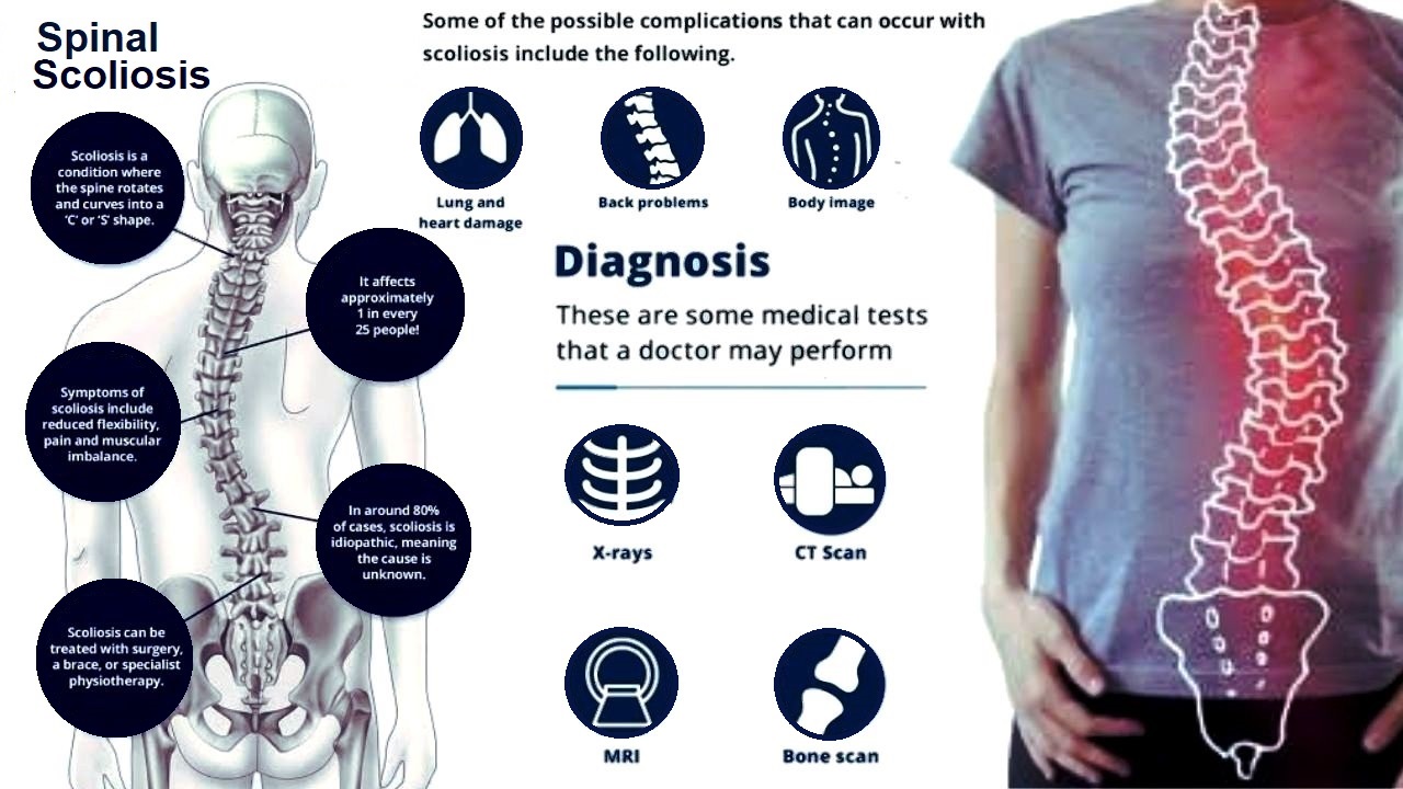
If degeneration is more pronounced on one side of the spine, degenerative scoliosis can result. The degenerative scoliosis curve, which is often located in the low back (lumbar spine), forms a slight "C" shape as the spine abnormally curves on one side or the other. Any sideways spinal curve of at least 10 degrees, as measured by the Cobb angle on spinal radiographs (X-rays), is considered scoliosis.
If degenerative scoliosis becomes symptomatic, pain can range from a dull back ache to excruciating sensations that shoot down the leg, commonly referred to as sciatica, and make walking difficult or impossible.Narrowing of the intervertebral disc space with sclerosis of the adjacent vertebral bodies may occur as a consequence of infection, neoplasia, trauma, or rheumatic disease. Some patients have been described with backache and these radiological appearances without any primary cause being apparent. The lesions were almost always of 1 or, at most, 2 vertebrae and most frequently involved the inferior margin of L4. We describe 3 patients with far more extensive vertebral involvement and present the clinical, radiological, scintiscan, and histological findings. The only patient we have seen with the better known, isolated L4/5 lesion was shown on biopsy to have staphylococcal osteomyelitis. For this reason we would still recommend a biopsy of all such sclerotic vertebral lesions if they occur in the absence of other rheumatic disease.