Labs and Tests
Your doctor may order blood tests to determine if you have inherited a blood disorder associated with DVT and PE. The blood tests are also used to measure carbon dioxide and oxygen levels. A blood clot in the lungs can lower oxygen levels in the blood.1
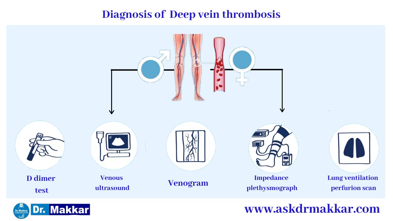
D-dimer test is usually used to rule out DVT in people with a low or intermediate risk for the condition. The test indicates whether you have elevated levels of D-dimer, a protein fragment that's left over from a clot once it's formed. If your D-dimer test is not elevated, chances are you do not have DVT.
Ultrasound
This is typically the preferred option for diagnosis. A venous ultrasound is usually done on people who have a history of DVT and are taking blood thinners and those who have a positive D-dimer test.
There are different types of venous ultrasonography:
Compression ultrasound (B-mode imaging): Similar to the duplex ultrasonography, compression ultrasound is a variation of the commonly-used medical ultrasound technique (also known as an “echo” test).
Duplex ultrasound (B-mode imaging and Doppler waveform analysis): Duplex ultrasonography uses high-frequency sound waves to visualize the flow of blood in the veins. It can detect blood clots in the deep veins and is one of the quickest, most painless, reliable, and noninvasive ways to diagnose DVT.2 The duplex ultrasonography also includes a color-flow Doppler analysis.
Color Doppler imaging: This produces a 2-D image of the blood vessels. With a Doppler analysis, a doctor can see the structure of the vessels, where the clot is located, and the blood flow. The Doppler ultrasound can also estimate how quickly blood is flowing and reveal where it slows down and stops. As the transducer is moved, it creates an image of the area.
The reliability of these tests varies. For example, compression ultrasounds are best for detecting DVT in proximal deep veins, like femoral and popliteal veins (thighs), but duplex ultrasound and color Doppler imaging are best for DVT of the calf and iliac veins (pelvis).
Venogram
In the past, making a firm diagnosis of DVT required performing a venogram. With a venogram, a contrast iodine-based dye is injected into a large vein in the foot or ankle, so doctors can see the veins in the legs and hips. X-ray images are made of the dye flowing through the veins toward the heart. This allows for doctors and medical professionals to see major obstructions to the leg vein.
This invasive test can be painful and entails certain risks, such as infection, so doctors generally prefer to use the duplex ultrasonography method.2
However, some doctors will use a venogram for people who have had a history of DVT. Because blood vessels and veins in these individuals are likely damaged from previous clots, a duplex ultrasonography won't be able to detect a new clot like a venogram can.
MRI and CT Scans
Magnetic resonance imaging (MRI) and computed tomography (CT) scans can create images of the organs and tissues in the body, as well as veins and clots. While useful, they are generally used in conjunction with other tests to diagnose DVT.
Lung Ventilation-Perfusion Scans; Pulmonary Angiography
If a CPTA isn't available, you might get a lung ventilation-perfusion scan or a pulmonary angiography.2
With the lung ventilation-perfusion scan, a radioactive substance shows the blood flow and oxygenation of the lungs. If you have a blood clot, the scan might show normal amounts of oxygen but slowed blood flow in parts of the lungs that have clotted vessels.
With a pulmonary angiography, a catheter from the groin injects a contrast dye into the blood vessels, which allows doctors to take X-ray images and follow the path of the dye to check for blockages.
Impedance Plethysmography
Impedance plethysmography is another non-invasive test for diagnosing DVT. While this test is reliable, many hospitals do not have the equipment or the expertise readily available to perform this test efficiently.
In impedance plethysmography, a cuff (similar to a blood pressure cuff) is placed around the thigh and inflated in order to compress the leg veins. The volume of the calf is then measured (by means of electrodes that are placed there). When the cuff deflates, it allows the blood that had been "trapped" in the calf to flow out th